Safety Rate
Cardiac Intravascular Ultrasound (IVUS) is an advanced imaging technique that provides a detailed view of the inside of your heart’s arteries. Unlike traditional angiography, which shows only the outline of blood vessels, IVUS uses high-frequency sound waves to reveal the artery’s inner structure. This helps cardiologists assess plaque buildup, vessel size, and blockage severity with remarkable accuracy. Cardiac Intravascular Ultrasound plays a crucial role in diagnosing complex heart conditions and planning precise treatments like angioplasty or stent placement.
Think of IVUS as a gentle window into your arteries — one that offers clarity, reassurance, and precision in protecting your heart health. This is especially true with advanced Intravascular Ultrasound (IVUS) now available in Chennai.
When a doctor needs to take a close look inside a heart artery, a regular X-ray or external scan is not always enough. This is where Intravascular Ultrasound becomes very useful. It gives a clear, detailed picture from inside the artery itself — something no outside scan can match.
Dr. Karthick Sabapathi is an experienced interventional cardiologist based in Chennai. He uses Intravascular Ultrasound as part of his approach to understanding and treating blocked or narrowed heart arteries. His focus is always on gathering the right information before making any treatment decision, so that every patient receives care suited to their specific condition.
Intravascular ultrasound — also written as IVUS — is a medical imaging technique used to look at the inside walls of the arteries that supply blood to the heart. The word "intravascular" simply means "inside the blood vessel."
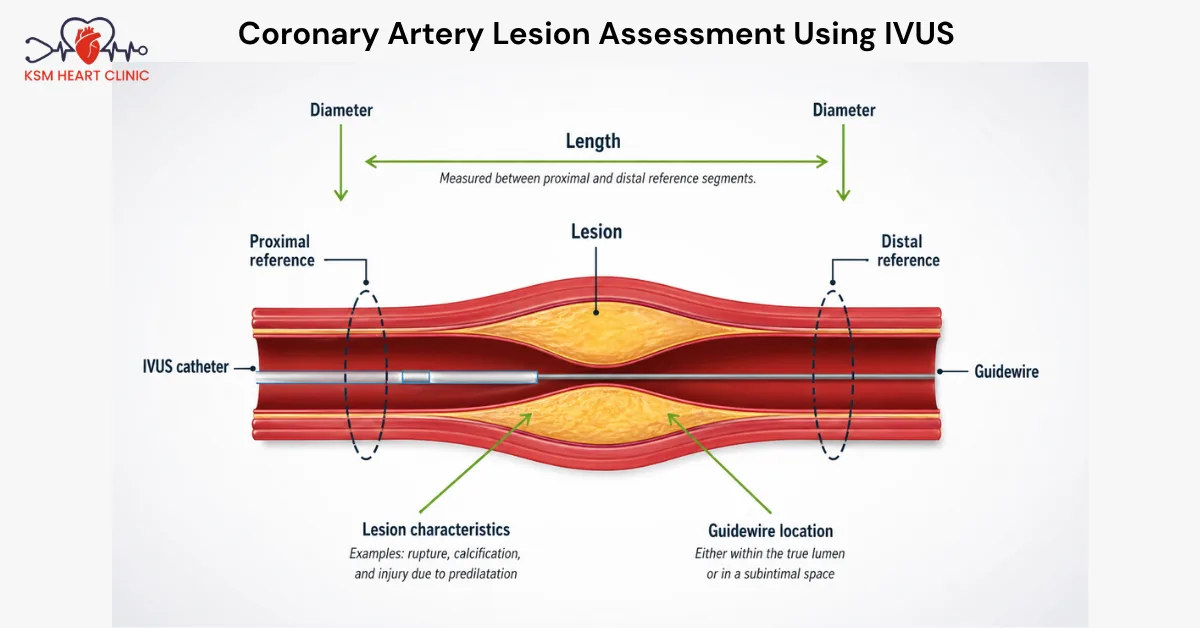
A very small ultrasound probe is placed at the tip of a thin, flexible tube called a catheter. This catheter is guided through the blood vessel until it reaches the area of concern. The probe then sends out sound waves. These sound waves bounce off the artery walls and return as signals, which are converted into real-time images on a monitor.
Unlike a standard angiogram, which shows only the shadow of blood flowing through the artery, intravascular ultrasound shows the actual thickness and condition of the artery wall. It can reveal plaque build-up, calcium deposits, and any weak spots in the vessel wall — details that are often invisible in a normal scan.
Dr. Karthick Sabapathi uses cardiac intravascular ultrasound to get a full picture of what is happening inside the artery before deciding on the best way to treat it.
The procedure begins like a standard cardiac catheterization. A small puncture is made, usually in the wrist or the groin area. A thin, flexible tube (catheter) is then carefully guided through the blood vessels towards the heart arteries using X-ray guidance.
At the tip of this catheter sits the ultrasound probe — no bigger than a few millimetres. Once it reaches the target area inside the artery, the probe begins rotating and sending out high-frequency sound waves in all directions.
As the catheter is slowly pulled back through the artery, it captures a continuous series of cross-sectional images — almost like taking slices of the artery at every millimetre. These images are displayed live on a screen and can be reviewed in detail.
The result is a precise, three-dimensional-like view of the inside of the artery — showing the lumen (the opening through which blood flows), the artery wall layers, and any blockages or abnormalities. This level of detail is what makes intravascular ultrasound so valuable in planning and delivering accurate cardiac treatment.

There are several ways this procedure adds value to your cardiac care:
In short, intravascular ultrasound treatment in Chennai under Dr. Karthick Sabapathi gives you the advantage of more accurate diagnosis and more precisely guided treatment — which leads to better outcomes.
Here is a step-by-step overview of what to expect:
You will be asked to avoid eating or drinking for a few hours before the procedure. Blood thinning medication may be adjusted beforehand. The care team will explain everything and answer any questions you have.
You will be given local anaesthesia at the access point — usually the wrist or groin — to numb the area. You will be awake throughout but made comfortable. A catheter is gently inserted and guided to the heart arteries. Once in position, the IVUS catheter captures detailed images while being slowly pulled back through the artery.
The imaging part of the procedure takes around 10 to 20 minutes. If stent placement or another intervention is being done at the same time, the total procedure time will be longer.
You will rest in a recovery area while the care team monitors your vital signs. Most patients are able to go home the same day or the following morning, depending on what was done. Dr. Karthick Sabapathi will discuss the findingsx with you and explain the next steps in your care.
There are clear, well-established reasons why cardiac intravascular ultrasound is considered a gold standard imaging tool in interventional cardiology:
These benefits make intravascular ultrasound treatment in Chennai a reliable choice for patients who want the most accurate and careful approach to their cardiac care.
Intravascular ultrasound is a well-established and generally safe procedure. It is performed routinely alongside coronary interventions and has a strong track record of safety.
That said, as with any invasive cardiac procedure, there are some small risks to be aware of:
It is important to note that the IVUS imaging probe itself does not use radiation — it uses sound waves, which are safe. The X-ray guidance used to position the catheter does involve a small amount of radiation, but this is carefully managed and kept to the minimum necessary.
Dr. Karthick Sabapathi will always discuss the specific risks and benefits with you before any procedure, taking into account your overall health and medical history. The goal is always to choose the safest and most effective path forward for each patient.
Intravascular ultrasound is one of the most powerful tools available in modern cardiac catheterisation. It gives cardiologists a level of detail about the inside of heart arteries that simply cannot be obtained any other way.
For patients in Chennai who need coronary artery evaluation or stent placement, Dr. Karthick Sabapathi brings the expertise and technology to make this procedure work to your advantage. With a careful, image-guided approach, he aims to ensure that every intervention is not just effective, but as safe and precise as possible.
The IVUS (Intravascular Ultrasound) cost in India generally ranges from ₹25,000 to ₹60,000, depending on the hospital, city, and whether it’s combined with procedures like angioplasty. The cost covers the imaging catheter, specialized equipment, and expert analysis to provide detailed insights into artery structure and plaque buildup.

IVUS is not always necessary with angioplasty but is often used to improve accuracy. It helps cardiologists visualize artery walls, assess blockage severity, and guide stent placement. While not mandatory, using IVUS during angioplasty enhances safety, optimizes stent positioning, and improves long-term treatment outcomes for patients with complex heart conditions.
During an IVUS procedure, a thin catheter with an ultrasound probe is inserted through an artery, usually via the wrist or groin, and guided to the heart. The probe emits sound waves, creating detailed images of the artery’s inner walls. This helps doctors assess blockages, vessel size, and plaque composition precisely.
Our experienced anaesthesiologists are here to ensure your safety and comfort